


Introduction
Oral cancer is cancer that develops in the tissues of the mouth or throat. It belongs to a larger group of cancers called head and neck cancers. Most develop in the squamous cells found in your mouth, tongue, and lips. Oral cancer is one of the most frequent cancers globally: in 2018, oral cancer occurred globally in about 355,000 people and resulted in 177,000 deaths, and unfortunately its late stage detection is a common problem worldwide. Early discovery is the most important step in reducing the death rate: when detected in the first stage, patients have an 80 to 90 percent survival rate.
Risk factors include:
- Smoke, the high concentration of carcinogenic substances contained in tobacco makes it very harmful and capable to irreversibly damaging the cells of the oral mucosa.
- Alcohol abuse, alcohol consumers have a 6 times higher risk than non-drinkers.
- Microtraumas from dental anomalies, altered dentitions or prostheses in poor condition (frequent in elderly subjects).
- Betel quid chewing, with or without added tobacco, increases the risk of oral/oropharyngeal cancer.
- Papillomavirus infection, with high oncogenic potential.
- It must be highlighted that 25% of oral cancer patients neither drink nor smoke.
Usually, oral cancer appears as a lesion, often ulcerated, painful, and easily bleeding, which spontaneously does not heal and can cause pain at rest. In other cases, it can appear with a later cervical lymph node swelling or with a mass of the lateral cervical region hard on palpation, not very mobile on the underlying strata, with intact skin, of increasing volume, and expression of local metastasis.
Knowing the risk factors and a regular examination, including a screening of the entire mouth, are essential in the early detection of cancerous and precancerous lesions.
The implementation of screening for early diagnosis is vital to reduce the morbidity and mortality associated with this pathology.
The ideal time to detect a tumour lesion and to have surgery is in the precancerous stage, where the prognosis for the patient is the best. Precancerous epithelial lesions typically begin below the tissue surface and grow to occupy the entire epithelium.
Staging of dysplasia
Different stages of dysplasia are recognised:
- Mild
- Moderate
- Severe
When dysplasia takes the entire epithelium, it is called carcinoma in situ and is classified as ‘invasive oral squamous cell carcinoma’ when it also occupies the basement membrane.
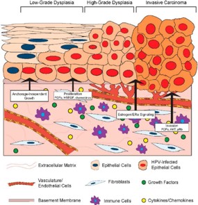
Fig. 1 Degrees of dysplasia in oral cancer
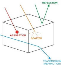
Fig. 2 The behavior of light in the presence of an object
Dysplasia and loss of autofluorescence
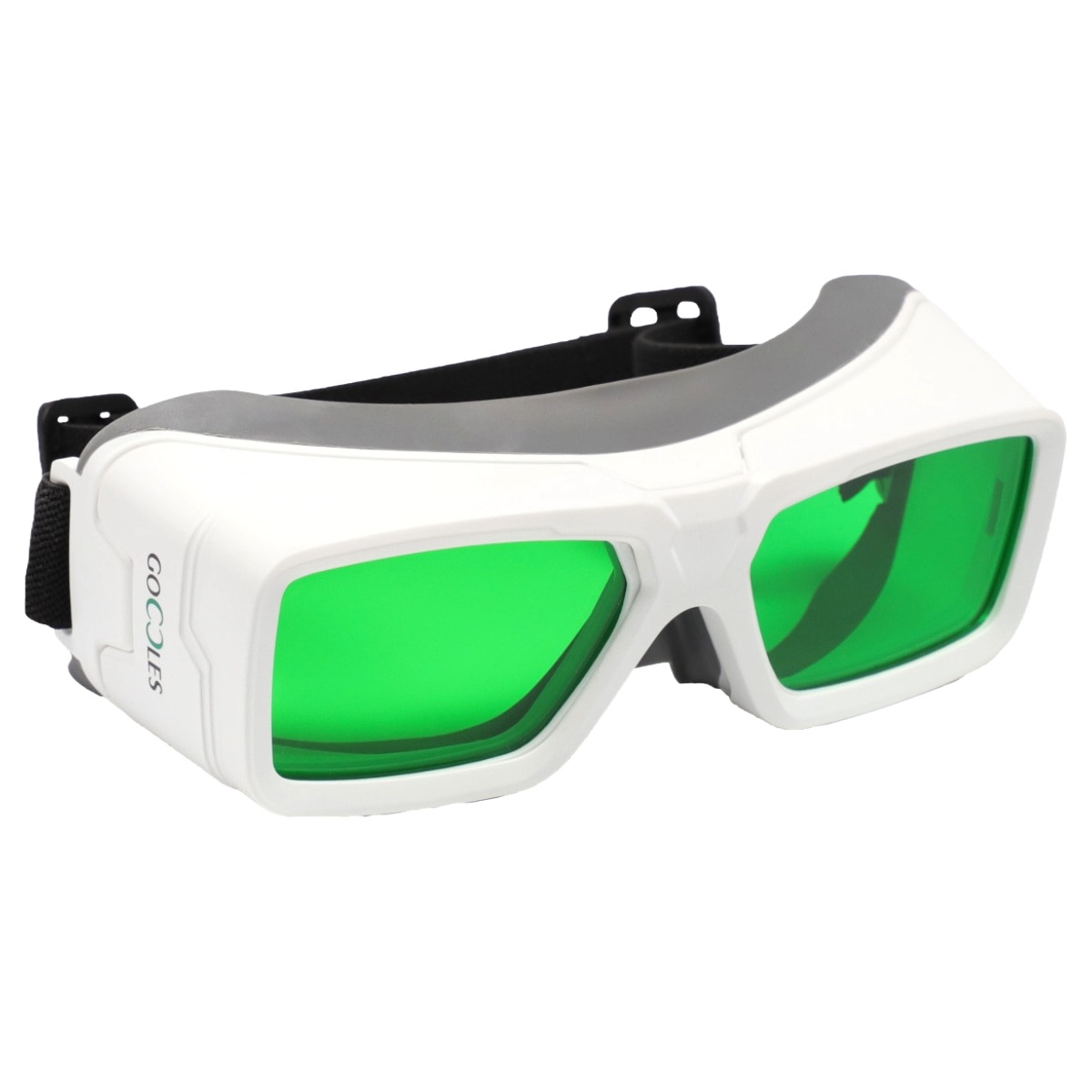
Let’s review, then, how dysplasia and tumours of the oral cavity can result in a decreased intensity of fluorescence in blue light and optical filter screening with the Goccles eyewear.
Essentially the loss of fluorescence in the presence of tumour processes is possible in 4 possible conditions:
1. The increased metabolic activity of dysplastic cells in the epithelium causes a decrease in FAD with a consequent reduction in fluorescence.
2. The breakdown of collagen matrices as a prelude to the advancement of cancer cells leads to a decrease in fluorescence
3. Morphological changes that can occur in dysplastic cells involve an increased scattering or scattering of light in the epithelial layer. This increases the backscattering of blue light which excites which excites the fluorescent component of the cells, with a consequent evident decrease in fluorescence (visible phenomenon even with the naked eye)
4. An increased intake of blood due to an increase of activity of dysplastic epithelium cells will result in a greater microvasculature of the stroma and therefore a greater absorption of light by the blood. Abnormal growth of dysplastic cells in the epithelium also leads to an increase in blood flow to the affected area as a normal physiological response. These two processes will result in a diminished fluorescence (phenomenon visible even with the naked eye).
The loss of fluorescence, however, is not only due to dysplasia or oral cancer, and this represents a limitation of this technique. Various causes can be associated with different types of lesions, as well as areas of prominent vascularisation, areas of inflammation and excessive melanin pigmentation, all attributable to a loss of fluorescence.
For this reason, in the presence of areas with loss of fluorescence, it is essential to follow the course over the next 2 to 4 weeks to observe the regression or persistence of the observed phenomenon.
Conclusions
Screening for oral cancer, using tissue fluorescence, is part of a clinical examination that includes the patient’s medical history with the risk assessment of the tumour, head-neck palpation and a first visual screening with the naked eye illuminated by white light.
In the presence of areas with loss of fluorescence, it is essential to follow the course over the next 2 to 4 weeks to observe the regression or persistence of the observed phenomenon.
The only way to accurately determine a diagnostic finding is with a biopsy and histopathological examination that allows identification of the possibility of a false positive. When performing tissue fluorescence screening, it may be helpful to have clinical imaging support from a pathologist or surgeon who has experience with this method, GOCCLES offers this support.

